Data Analysis
Statistical consultation is available through the HIMC Statistical Consulting Service.
The Human Immune Monitoring Center (HIMC)
Immunoassays
At Stanford HIMC, we offer standardized immunoassays for human and mouse cytokines, for analysis of serum, plasma, cell culture supernatants, tissue homogenates and other fluids. We measure multiple cytokines simultaneously using Luminex Bead Array or Olink Target 96 Arrays (which utilizes PEA Technology), and Alamar NULISA assay (which utilizes PLA technology).
- Human 80-plex cytokine panel
- Human 48-plex cytokine panel (=Panel 1 of 80-plex)
- Mouse 48-plex cytokine panel
- Mouse 64-plex
- Custom panels (e.g., from ThermoFisher, and others)
Biomarker information in our standard Luminex kits can be downloaded here.
Olink Panels
- Target 96 panels: Inflammation, Cardiometabolic, Cardiovascular II, Cardiovascular III, Immuno-oncology, Immune response, Oncology, Oncology II, Oncology III, Neurology, Neuro-exploratory, Organ Damage, Development, Metabolism, Cell Regulation, Mouse Exploratory
- Target 48 panels: Human cytokine, Mouse cytokine
Alamar NULISA Panels
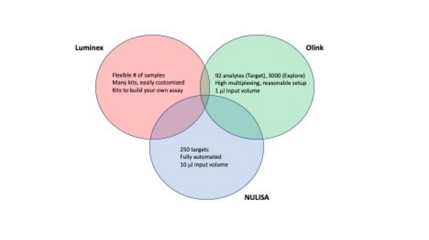
How to choose between platforms?
To decide between the different platforms, we have resources to guide you through your selection. Please don’t hesitate to contact by emailing: maecker@stanford.edu .
Here below is a short overview of the Luminex, Olink and NULISA assays
Additionally, for questions regarding your choice of Luminex vs. Olink, see also our most recent FAQ’s here.
User Instructions
1. Planning your assay:
We suggest analyzing all samples in a single experiment, to minimize run to run variability. To account for plate-to-plate differences and downstream analysis, it is recommended to evenly distribute groups across multiple plates (i.e., don't place all controls on one plate and all test samples on another). However, all time points per subject should be in the same plate. Samples will be run in the order provided, unless instructed otherwise. Please consult with us for optimized design and sample distribution in the planning phase of your experiments.
2. Collecting your samples:
For Human 80 plex we request a minimum of 200 microliters per sample to run duplicate wells without dilution. Be sure samples are processed and spun before freezing, to remove cells and debris that can clog the Luminex instrument; see our SERUM PROCESSING or PLASMA PROCESSING protocols for details on serum or plasma collection and processing. Process serum and plasma within two hours of blood draw if possible. Avoid hemolysis, which interferes with cytokine detection. Serum and plasma processing services are also available through https://med.stanford.edu/ctru.html. For mouse and rodent samples standard processing vials are available: Kent Scientific & Mini Collect , we request 50-70ul of plasma or serum samples . For Tissue homogenates please include a buffer with protease inhibitors, and normalize samples to total protein. For cell supernatants include media with serum as background. If questions, please email: maecker@stanford.edu.
All samples should be frozen at -80C and transferred to the HIMC on dry ice. Multiple freeze-thaw cycles should be avoided. Please label all tubes clearly on the side and top, email an excel list of your samples to maecker@stanford.edu (or post to iLab or Basecamp for existing projects).
3. Running samples:
An online order is required before samples can be run. Turnaround time varies by the project size (number of samples) and the current schedule; Projects with fewer than 20 samples can be completed within two weeks of order and sample delivery. We are located at: 1651 Page Mill Rd. room 0280, 650-723-4984.
Plasma and Serum samples are run with a 1:3 dilution which is optimal for these matrices and prevents clogging of the instrument; dilution of other sample types is optional; it can improve quantitation of high-level cytokines, with minimal impact on the detection of most low-level cytokines. Leftovers will be discarded unless you provide instructions and will not be saved for more than two weeks.
4. Data analysis:
Data is shared via our Basecamp project management system. New customers will receive an email invitation to join the system. An Excel report will be posted there, containing summaries of median fluorescence intensity (MFI), calculated concentration, bead count, and coefficient of variation (CV) of replicate wells, for each cytokine in each sample, standard, and control. QC notes are also provided in the report. The data is simultaneously uploaded to our online database, Stanford Data Miner, where it can be integrated with other HIMC data from your lab, as well as clinical/demographic data if provided. Click the help link in the upper left corner of the Data Miner homepage for instructions on using this system.
In general, for Luminex assays, the HIMC recommends analyzing raw data (median fluorescence intensity) rather than concentration (pg/mL). Reasons for this recommendation are many and complex. Most prominent is that standard curves are generated in a matrix that differs from biological matrix of your specimens so, in this sense, standard curves can be biased. Also, standard curves become non-linear at high and low ends, yielding unreliable and biased concentrations.
Normalization (to controls or to global sample median for each plate) is recommended when comparing values across plates.
Charges:
Regarding costs, see the Prices and Ordering tab in the left menu for instructions on ordering services. Please note that Olink assays are charged per array rather than per sample, so it’s best if you have close to a full array of samples (88 plus our required controls).
Additional information:
Methods for collection mouse blood
Protocols
See the Protocols page for complete HIMC protocols as well as short descriptions suitable for publications and grant applications.
Statistical consultation is available through the HIMC Statistical Consulting Service.
For more information
Contact:
Holden Maecker, HIMC Director
Address:
Human Immune Monitoring Center
1651 Page Mill Road, Lab 0280, Palo Alto, CA 94034-5518
phone: 650-723-1671
email: maecker@stanford.edu